
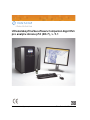
Companion Algorithm p53 (DO-7) Image Analysis Software User Manual, v. 5.1
p53 (DO-7) Handbuch zur Bildanalyse-Software-Anwendung, v. 5.1
Manuel d’utilisation de l’application logicielle d’analyse d’images
du p53 (DO-7), v. 5.1
Manual del usuario de la aplicación del software de análisis de imagen p53
(DO-7), v. 5.1
Manuale dell’utente del software per l’analisi delle immagini di
p53 (DO-7), v. 5.1
Uživatelská příručka softwarové aplikace pro analýzu obrazu
p53 (DO-7), v. 5.1
Brugervejledning for Softwareapplikation til p53 (DO-7) billedanalyse, v. 5.1
Εγχειρίδιο χρήσης εφαρμογής του λογισμικού ανάλυσης εικόνας της p53
(DO-7), v. 5.1
p53 (DO-7) képelemző szoftveralkalmazás használati útmutató, v. 5.1
Gebruikershandleiding voor de p53 (DO-7) beeldanalyse-softwareapplicatie,
v. 5.1
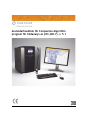
Brukerhåndbok for p53 (DO-7) bildeanalyseprogram, v. 5.1
Podręcznik użytkownika oprogramowania użytkowego do analizy obrazu
p53 (DO-7), v. 5.1
Software de análise de imagem de p53 (DO-7), v. 5.1
Priročnik za uporabo programske opreme za analizo slik p53 (DO-7), v. 5.1
Användarhandbok för programmet för bildanalys av p53 (DO-7), v. 5.1
Not for distribution in the US
PS-001603/06449042001
Ventana Medical Systems, Inc. Part No.: 101407300 Rev. A

INTENTIONALLY BLANK

Companion Algorithm p53 (DO-7) Image Analysis
Software User Manual, v. 5.1
Ventana Medical Systems, Inc. Part No.: 101407300 Rev. A

Companion Algorithm p53 (DO-7) Image Analysis Software User Manual
Copyright
Copyright © 2014. Ventana Medical Systems, Inc. All rights reserved.
Trademarks
BENCHMARK, CONFIRM, COMPANION ALGORITHM, VENTANA ISCAN HT, ISCAN, VIRTUOSO,
VENTANA, and the VENTANA logo are trademarks of Roche. All other trademarks are the property of their respective
owners.
Companion Algorithm p53 (DO-7) image analysis software is licensed for use between Ventana Medical Systems, Inc.
and a licensee, and only users authorized there under are permitted to access and use the software. Unauthorized use and
distribution may result in civil and criminal penalties.
Open Source and Commercial Software
Refer to the Virtuoso Reference Guide for information on Open Source and Commercial Software programs.
Contact Information:
Ventana Medical Systems, Inc.
1910 E. Innovation Park Drive
Tucson, AZ 85755
USA
+1 520 887 2155
www.ventana.com
Ventana Medical Systems, Inc.
203 Ravendale Drive
Mountain View, CA 94043
USA
Roche Diagnostics GmbH
Sandhofer Strasse 116
D-68305 Mannheim
Germany
+49 621 7590

Part No.: 101407300 Rev. A i
Table of Contents
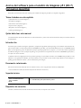
About the Companion Algorithm p53 (DO-7) image analysis software . . . . . . . . . .1
Topics Covered in this Chapter . . . . . . . . . . . . . . . . . . . . . . . . . . . . . . . . . . . . . . . . . . . 1
Who Should Read this Manual . . . . . . . . . . . . . . . . . . . . . . . . . . . . . . . . . . . . . . . . . . . . 1
Introduction . . . . . . . . . . . . . . . . . . . . . . . . . . . . . . . . . . . . . . . . . . . . . . . . . . . . . . . 1
Related Document . . . . . . . . . . . . . . . . . . . . . . . . . . . . . . . . . . . . . . . . . . . . . . . . . . . 1
Technical Support. . . . . . . . . . . . . . . . . . . . . . . . . . . . . . . . . . . . . . . . . . . . . . . . . . . . 1
Connection Requirements . . . . . . . . . . . . . . . . . . . . . . . . . . . . . . . . . . . . . . . . . . . . . . 1
Cyber Security . . . . . . . . . . . . . . . . . . . . . . . . . . . . . . . . . . . . . . . . . . . . . . . . . . . . . . 2
User Authorization . . . . . . . . . . . . . . . . . . . . . . . . . . . . . . . . . . . . . . . . . . . . . . . . . 2
Securing Networks and Servers. . . . . . . . . . . . . . . . . . . . . . . . . . . . . . . . . . . . . . . . . 2
Protecting Data . . . . . . . . . . . . . . . . . . . . . . . . . . . . . . . . . . . . . . . . . . . . . . . . . . . 2
Chapter 1: Intended Use and Indications for Use . . . . . . . . . . . . . . . . . . . . . . . .5
Intended Use and Indications for Use . . . . . . . . . . . . . . . . . . . . . . . . . . . . . . . . . . . . . . . 5
Summary and Explanation . . . . . . . . . . . . . . . . . . . . . . . . . . . . . . . . . . . . . . . . . . . . 5
Test Principles . . . . . . . . . . . . . . . . . . . . . . . . . . . . . . . . . . . . . . . . . . . . . . . . . . . . . . 6
Warnings and Precautions . . . . . . . . . . . . . . . . . . . . . . . . . . . . . . . . . . . . . . . . . . . . 6
Pre-Analytical Variables . . . . . . . . . . . . . . . . . . . . . . . . . . . . . . . . . . . . . . . . . . . . . 6
Procedure . . . . . . . . . . . . . . . . . . . . . . . . . . . . . . . . . . . . . . . . . . . . . . . . . . . . . . 6
Required Materials Not Provided. . . . . . . . . . . . . . . . . . . . . . . . . . . . . . . . . . . . . . . . 6
Results . . . . . . . . . . . . . . . . . . . . . . . . . . . . . . . . . . . . . . . . . . . . . . . . . . . . . . . . . . . 6
Limitations . . . . . . . . . . . . . . . . . . . . . . . . . . . . . . . . . . . . . . . . . . . . . . . . . . . . . . 6
Performance Characteristics . . . . . . . . . . . . . . . . . . . . . . . . . . . . . . . . . . . . . . . . . . 7
Assay Cutoff . . . . . . . . . . . . . . . . . . . . . . . . . . . . . . . . . . . . . . . . . . . . . . . . . . . . . 7
Chapter 2: p53 Comparison and Reproducibility Studies . . . . . . . . . . . . . . . . . . .9
p53 Marker Studies . . . . . . . . . . . . . . . . . . . . . . . . . . . . . . . . . . . . . . . . . . . . . . . . . . . 9
Staining Procedure. . . . . . . . . . . . . . . . . . . . . . . . . . . . . . . . . . . . . . . . . . . . . . . . . 9
Study Devices and Samples . . . . . . . . . . . . . . . . . . . . . . . . . . . . . . . . . . . . . . . . . . . 9
p53 Concordance with BenchMark XT Instrument and iScan Coreo Slide Scanner . . . . . . . . 9
Intra-System and Inter-System Studies. . . . . . . . . . . . . . . . . . . . . . . . . . . . . . . . . . . 10
P53 Concordance with BenchMark ULTRA Instrument and iScan Coreo Slide Scanner. . . . . 12
p53 Concordance with BenchMark XT/ULTRA Instruments and VENTANA iScan HT Slide Scanner13
p53 Concordance with BenchMark GX Instrument and iScan Coreo and iScan HT Slide Scanners14
Appendix A: Reagents (Antibody) Package Inserts. . . . . . . . . . . . . . . . . . . . . . .19
Reagents (Antibody) Package Inserts . . . . . . . . . . . . . . . . . . . . . . . . . . . . . . . . . . . . . . 19
Index . . . . . . . . . . . . . . . . . . . . . . . . . . . . . . . . . . . . . . . . . . . . . . . . . . . . .21

ii Part No.: 101407300 Rev. A
Table of Contents p53 (DO-7) Image Analysis Software User Manual
INTENTIONALLY BLANK

Part No.: 101407300 Rev. A 1
About the Companion Algorithm p53 (DO-7) image analysis
software
Welcome to the Companion Algorithm p53 (DO-7) Image Analysis Software User Manual.
Topics Covered in this Chapter
• Who Should Read this Manual (page 1)
• Introduction (page 1)
• Related Document (page 1)
• Technical Support (page 1)
• Connection Requirements (page 1)
• Cyber Security (page 2)
Who Should Read this Manual
System Administrators should read this user manual and use it for reference while operating the VENTANA Virtuoso software.
Introduction
The Companion Algorithm p53 (DO-7) image analysis software assists the pathologist in the semi-quantitative measurement of p53
in tissues stained with Ventana Medical Systems, Inc. CONFIRM anti-p53 (DO-7) Primary Antibody (CONFIRM anti-p53 (DO-7)).
This application generates a p53 score that can be reviewed and accepted by the pathologist, or if necessary, overridden by the
pathologist. The image analysis application is an assist to the pathologist in the scoring and interpretation of CONFIRM anti-p53
(DO-7) staining on breast cancer tissues.
The Virtuoso p53 Digital Read Application allows the pathologist to view CONFIRM anti-p53 (DO-7) stained slides as images on
a computer monitor, similar to what can be viewed under a microscope. While reviewing the image, the pathologist may change
magnification and move freely about the image.
Related Document
For additional information on the Virtuoso software, see the following VENTANA document:
• Virtuoso Reference Guide (PL-000123-EN)
Technical Support
Connection Requirements
Refer to the Virtuoso Reference Guide information on connection requirements.
Ventana Medical Systems, Inc.
1910 E. Innovation Park Drive
Tucson, AZ 85755
USA
Tel: +1 520 887 2155; for Technical Support, press 1

2 Part No.: 101407300 Rev. A
About the Companion Algorithm p53 (DO-7) image analysis software p53 (DO-7) Image Analysis Software User Manual
Cyber Security
Any device that is connected to a network (internally or externally) has the potential to be compromised by unauthorized access or
viruses. As with most devices, the software is designed to run on a computer utilizing Microsoft Windows and virus protection
software which requires the validation and implementation of the appropriate patches.
Some of the potential cyber security hazards are:
• Malicious software that alters the device software (such as viruses)
• Unauthorized access to the system that could compromise data safety
• Security of data transmitted over the Internet
Cyber security involves protecting data by preventing, detecting, and responding to malicious cyber attacks. Cyber attacks could
involve computer viruses which can completely erase data or hackers who alter files or even use the device as a host to attack other
devices. As serious as these hazards are, steps can be taken to maximize cyber security.
User Authorization
All software users must login with a valid user name and password. The user name and password are securely transmitted in
encrypted form over the Internet or Intranet. Once a user has logged in, the user remains active in the application until the user
explicitly logs out, closes the browser, or because the application closes after a period of inactivity.
Securing Networks and Servers
Network security consists of the provisions made in the computer network infrastructure, policies adopted by the network
administrator to protect the network, and the network resources that prevent unauthorized access.
The following are critical steps for securing a network server:
• Physical security (servers and network infrastructure behind locked doors)
• Use of robust passwords
• System and data backups (at regular intervals)
• Data protection
• Terminating unused services
• Restricting access to used services
The following are critical steps and methodologies used to secure network and servers:
• Data protection
• Data backups (at regular intervals)
• Refusal of automatic updates from off-the-shelf software
• Antivirus software for computers and servers
Protecting Data
Establishment of a network firewall and protection of the network against viruses using anti-virus software are effective methods to
protect data. Virus definitions should be kept up to date and regular scans of computers for spyware should be performed using a
legitimate anti-spyware application. If viruses or spyware are found, remove them immediately.

p53 (DO-7) Image Analysis Software User Manual About the Companion Algorithm p53 (DO-7) image analysis software
Part No.: 101407300 Rev. A 3
Evaluate Your Software Settings
The default settings of most software enable all available functionality. However, hackers may be able to take advantage of this
functionality to access devices. It is especially important to check the settings for software that connects to the Internet (browsers,
email clients, etc.). Apply the highest level of security available that still provides needed functionality.
Backup and Recovery
In order to develop a successful backup and recovery plan, comprehension of data accessibility needs and the potential impact of
data loss is essential. Automatic backup procedures need to be adopted using a data backup utility.

4 Part No.: 101407300 Rev. A
About the Companion Algorithm p53 (DO-7) image analysis software p53 (DO-7) Image Analysis Software User Manual
INTENTIONALLY BLANK

Part No.: 101407300 Rev. A 5
Chapter 1: Intended Use and Indications for Use
This chapter shows comparison and reproducibility studies for the p53 marker.
Intended Use and Indications for Use
The Virtuoso system provides automated digital slide creation, management, analysis, and viewing. It is intended for in vitro
diagnostic use as an aid to the pathologist in the display, detection, counting, review and classification of tissues and cells of clinical
interest based on particular morphology, color, intensity, size, pattern and shape.
Normal p53 protein acts as a tumor suppressor. Mutations in p53 may eliminate the tumor suppressive functions and allow
expression of malignancies. The IHC p53 (DO-7) Digital Read and Image Analysis applications are intended for use as an aid to the
pathologist in the detection and quantitative measurement of p53 protein in formalin-fixed, paraffin-embedded normal and
neoplastic tissue. When used with Ventana Medical Systems, Inc. CONFIRM anti-p53 (DO-7) Primary Antibody, it is indicated for
use as an aid in the assessment p53 protein of breast cancer patients (but is not the sole basis for treatment).
Note: The IHC p53 (D0-7) Digital Read and Image Analysis applications are adjunctive computer-assisted methodologies for the
qualified pathologist in the acquisition and measurement of images from microscope glass slides of breast cancer specimens
stained for the presence of p53 protein. The pathologist should verify agreement with the Image Analysis software application
score. The accuracy of the test results depends on the quality of the immunohistochemical staining. It is the responsibility of
a qualified pathologist to employ appropriate morphological studies and controls as specified in the instructions for the
CONFIRM anti-p53 (DO-7) Primary Antibody assay used to assure the validity of the Virtuoso System for IHC p53 Digital Read
and Image Analysis scores. The actual correlation of CONFIRM anti-p53 (DO-7) Primary Antibody to clinical outcome has not
been established.
Summary and Explanation
The Virtuoso system for p53 (DO-7) is an instrument and software system designed to assist the qualified pathologist in the
consistent quantitative assessment of protein expression in immunohistochemically (IHC) stained histologic sections from
formalin-fixed, paraffin-embedded (FFPE) normal and neoplastic tissues. The Virtuoso system can be used for review of digitized
images of histologic sections with image analysis algorithms (Companion Algorithm image analysis applications), or without
Companion Algorithm image analysis algorithms (Virtuoso Digital Read applications).
Digital Read applications present images on the computer screen in the same manner as one would see with a manual microscope,
inclusive of the pathologist's ability to select any areas of interest and the option of various magnification levels. For the Companion
Algorithm image analysis applications, the pathologist may use the system software to select and outline one or several field of views,
(FOVs), and each FOV may be viewed at various magnifications and then analyzed by the software; a count of the total number of
target cells and the number interpreted by the algorithm as positive and negative is generated. The pathologist can accept the score
provided by the algorithm, or may override the score with a pathologist score. The system requires competent human intervention
at all steps in the analysis process, and the software makes no independent interpretations of the data.
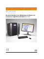
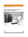
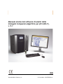
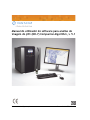
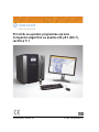
The Virtuoso system consists of a slide scanner, computer, monitor, keyboard, mouse, Companion Algorithm image analysis
algorithms, and software with a user interface. Virtuoso is an end-to-end digital pathology software solution that allows pathology
laboratories to acquire, manage, view, analyze, share, and report on digital images of pathology specimens. Using the Virtuoso
software, the pathologist can view digital images at various magnifications (as previously stated), add annotations, make
measurements, perform image analysis, and generate reports.
For in vitro diagnostic (IVD) use.

6 Part No.: 101407300 Rev. A
Chapter 1: Intended Use and Indications for Use p53 (DO-7) Image Analysis Software User Manual
Test Principles
The Virtuoso System for p53 (DO-7) employs image analysis techniques to obtain p53 scoring. Pre-defined parameters are used to
obtain p53 scores. The identification of the nucleus is carried out automatically by the image analysis algorithms. The steps involved
in the analysis algorithms are:
1. Enhancing the image. This process increases the contrast to make the image more suitable for analysis.
2. Identifying the epithelial area. The epithelial area is the region of the image where there is the possibility of epithelial cells
being present.
3. Identifying the nucleus.
4. Classifying the cells based on extent, intensity, and thickness of nuclear staining.
5. Computing the score.
Warnings and Precautions
It is important that glass slides with acceptable staining quality be used.
Pre-Analytical Variables
Tissue preparation and staining should follow the recommendations provided in the CONFIRM anti-p53 (DO-7) assay package
insert. For optimal image capture using the Virtuoso software, it is recommended that the tissue be free of folds and be placed on
the slide with a minimum of 2 mm boundary from the edge on all sides. The cover slip and slide label (if present) should not overhang
the edges of the slide. For further information on scanning, please refer to the appropriate iScan slide scanner reference guide.
Procedure
Refer to the Virtuoso Reference Guide.
It is recommended that at least three field of view be selected for analysis when using the Companion Algorithm p53 (DO-7)
software.
Required Materials Not Provided
The Virtuoso System for p53 DO-7 requires use of CONFIRM anti-p53 (DO-7), and any additional material or supplies listed in
the Ventana package insert, to stain tissues prior to analysis.
The iScan Coreo slide scanner or the VENTANA iScan HT slide scanner is required for scanning of the slides.
Results
The Virtuoso System for p53 DO-7 produces images and a staining score. The pathologist views the image and the instrument score,
makes an assessment, and reports a score which may not be the same as the instrument score. Refer to Virtuoso Reference Guide
for an example of a report.
Limitations
The algorithms are designed to work for p53 cell nuclei staining. The test results are only as good as the quality and accuracy of the
immunohistochemistry slide that is imaged, and the subsequent image that is analyzed. The pathologist must validate the p53 staining
run by examination of the p53 control images to verify that the expected results have been obtained before images from patient

p53 (DO-7) Image Analysis Software User Manual Chapter 1: Intended Use and Indications for Use
Part No.: 101407300 Rev. A 7
slides are analyzed. The pathologist must follow the manufacturer's recommendations for CONFIRM anti-p53 (DO-7) including
using all the positive and negative quality control materials for each staining run. If the control slides are not acceptable, the patient
tissues need to be re-stained with acceptable results. (See the Ventana CONFIRM anti-p53 (DO-7) package insert for details about
quality control recommendations.) The pathologist must follow the Ventana recommendations for surveying the entire breast
cancer specimen to assess any heterogeneity in the p53 staining, the degree of background staining, cytoplasmic staining, edge effect,
etc. as recommended in the Ventana CONFIRM anti-p53 (DO-7) user manual (available at www.ventana.com). If the images
captured have different staining (nuclear, cytoplasm, etc.), incorrect results will be generated. Incorrect results will also be generated
if the image quality cannot be analyzed. The software algorithms determine whether the quality of an image can be analyzed, based
on pre-defined parameters. Refer to the Virtuoso Reference Guide for more information.
The p53 (DO-7) algorithm will reject nuclei that are elongated regardless of the overall shape of the cell. For this reason, tumors
containing large numbers of cells with elongated nuclei may need to be evaluated manually. In addition, performance of the Virtuoso
System with the following types of breast cancers has not been evaluated: carcinoma in situ, carcinosarcoma, comedo carcinoma,
cystosarcoma phylloides, medullary carcinoma of the breast, mucinous variants of breast cancer, and spindle cell carcinoma.
Performance Characteristics
Performance of the staining agent is described in the Ventana package insert for the CONFIRM anti-p53 (DO-7) staining process.
See Comparison and Reproducibility Studies in Chapter 2 for a description of the performance of the software.
Assay Cutoff
Clinical cutoffs used for the assessment of p53 varies between laboratories. The performance of the Virtuoso system with p53
(DO-7) was evaluated at these commonly used clinical cutoffs: 0-0.99% was considered a negative test result and 1-10% and >10%
were considered positive test results. In some studies, the performance was evaluated at another commonly used clinical cutoff:
0-10% was considered a negative test result and >10% were considered positive test results.

8 Part No.: 101407300 Rev. A
Chapter 1: Intended Use and Indications for Use p53 (DO-7) Image Analysis Software User Manual
INTENTIONALLY BLANK
Part No.: 101407300 Rev. A 9
Chapter 2: p53 Comparison and Reproducibility Studies
This chapter shows comparison and reproducibility studies for the p53 marker.
p53 Marker Studies
Staining Procedure
Refer to the CONFIRM anti-p53 (DO-7) package insert for the BenchMark XT, ULTRA andGX instruments, ultraView, and iView
detection.
Study Devices and Samples
The p53 comparison and reproducibility studies for the Virtuoso Digital Read and Companion Algorithm image analysis software
consisted of 120 de-identified archived breast carcinoma sections immunohistochemically stained with CONFIRM anti-p53 (DO-7)
Primary Antibody. Study test samples covered the ranges of 0-0.99%, 1-10% and >10%, and were interpreted at three different sites
by three different pathologists. All test slides were scanned at 20X magnification and all images were output in bif file format.
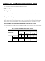
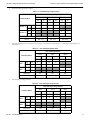
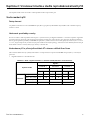
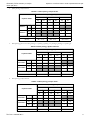
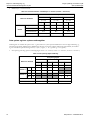
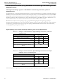
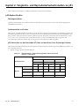
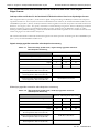
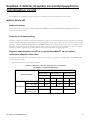
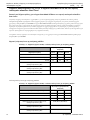
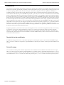
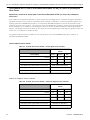
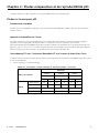
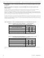
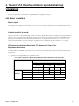
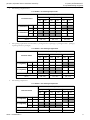
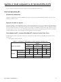
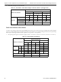
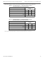
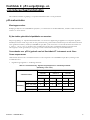
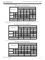
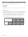
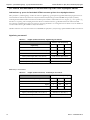
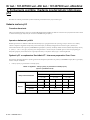
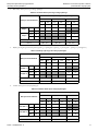
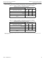
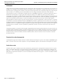
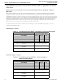
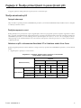
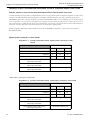
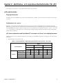
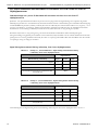
p53 Concordance with BenchMark XT Instrument and iScan Coreo Slide Scanner
The table below shows the concordance results for CONFIRM anti-p53 (DO-7) staining interpretation among three different sites:
1. Digital Read vs. Manual Method.
Table 2-1 Agreement - Digital Read vs. Manual (manual = true score).
Confusion Matrix
Digital
Site 1 Site 2 Site 3
(n = 119) (n = 119) (n = 118)
Neg Pos Neg Pos Neg Pos
Manual
Neg (0-0.99%) 24 8 53 12 41 8
Pos (1-10%, >10%) 9 78 4 50 11 58
% Agreement 86% 87% 84%
(95% CI) (78% - 91%) (79% - 92%) (76% - 89%)

10 Part No.: 101407300 Rev. A
Chapter 2: p53 Comparison and Reproducibility Studies p53 (DO-7) Image Analysis Software User Manual
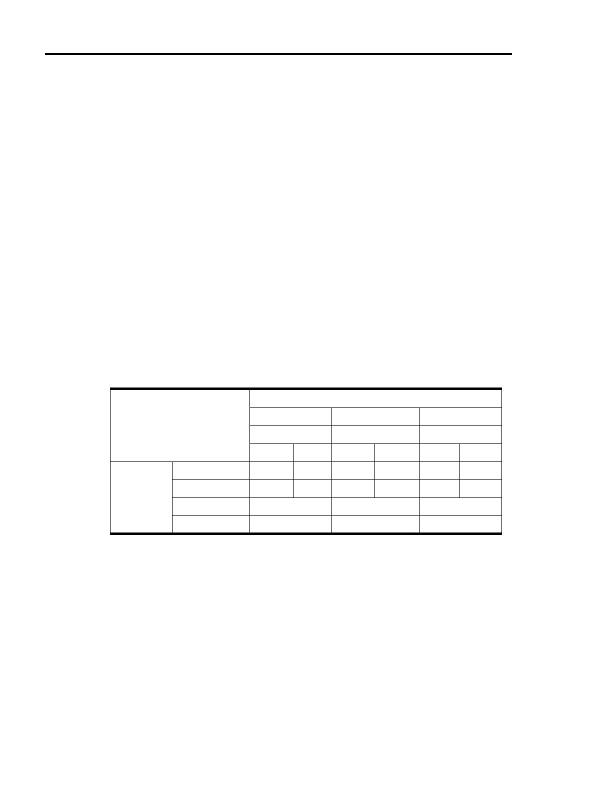
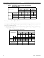
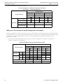
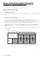
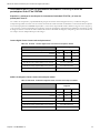
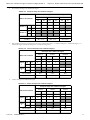
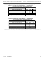
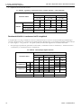
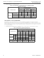
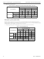
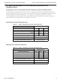
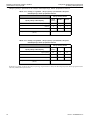
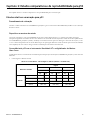
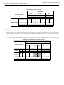
2. Image Analysis vs. Manual Method.
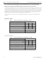
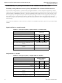
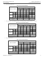
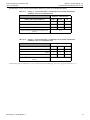
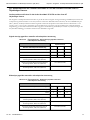
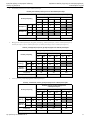
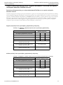
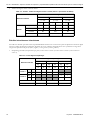
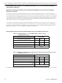
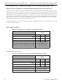
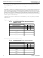
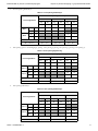
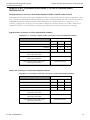
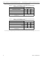
Intra-System and Inter-System Studies
The study was designed to demonstrate inter- and intra-Virtuoso system reproducibility for Virtuoso Digital Read and Companion
Algorithm image analysis applications. A designated subset of 40 cases that span the range of the p53 scoring categories (0-0.99%,
negative, 1-10%, positive, and >10%, positive) were used.
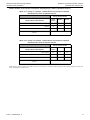
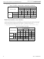
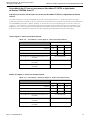
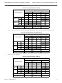
1. Intra-pathologist/Inter-Day (pair-wise comparisons, Session1 vs. Session 2, Session1 vs. Session 3, Session 2 vs. Session 3).
Table 2-2 Agreement - Image Analysis vs. Manual (manual = true score).
Confusion Matrix
Image Analysis
Site 1 Site 2 Site 3
(n = 119) (n = 119) (n = 118)
Neg Pos Neg Pos Neg Pos
Manual
Neg (0-0.99%) 16 16 46 19 33 15
Pos (1-10%, >10%) 0 87 1 53 5 65
% Agreement 87% 83% 83%
(95% CI) (79% - 92%) (75% - 89%) (75% - 89%)
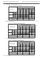
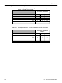
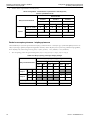
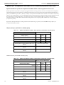
Table 2-3 Intra- Pathologist Digital Read.
Confusion Matrix
Intra-Pathologist Digital
Session 2 Session 3 Session 3
Neg Pos Neg Pos Neg Pos
10 30 10 30 10 30
Session 1
Neg 12 10 2 9 3
Pos 28 0 28 1 27
Session 2
Neg 10 91
Pos 30 129
% Agreement 95% 90% 95%
(95% CI) (83% - 99%) (77% - 96%) (83% - 99%)

p53 (DO-7) Image Analysis Software User Manual Chapter 2: p53 Comparison and Reproducibility Studies
Part No.: 101407300 Rev. A 11
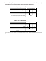
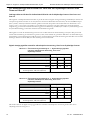
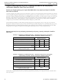
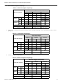
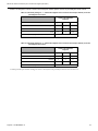
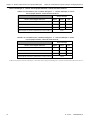
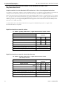
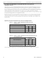
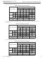
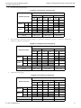
2. For Intra-Pathologist Image Analysis.
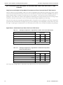
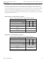
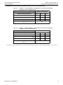
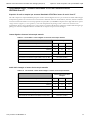
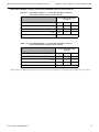
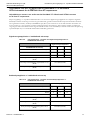
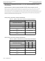
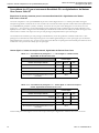
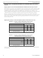
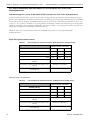
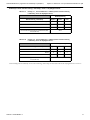
3. Inter-pathologist (pair-wise comparisons, Pathologist 1 vs. Pathologist 2, Pathologist 1 vs. Pathologist 3, Pathologist 2 vs.
Pathologist 3).
4. Inter-Pathologist Image Analysis.
Table 2-4 Intra-Pathologist Image Analysis.
Confusion Matrix
Intra-Pathologist Image Analysis
Session 2 Session 3 Session 3
Neg Pos Neg Pos Neg Pos
13 27 13 27 13 27
Session 1
Neg 12 9 3 11 1
Pos 28 4 24 2 26
Session 2
Neg 13 94
Pos 27 423
% Agreement 83% 93% 80%
(95% CI) (68% - 91%) (80% - 97%) (65% - 90%)
Table 2-5 Inter-Pathologist Digital Read.
Confusion Matrix
Inter-Pathologist Digital
Site 2 Site 3 Site 3
Neg Pos Neg Pos Neg Pos
57 62 52 66 52 66
Site 1
Neg 33 31 2 30 3
Pos 86 26 60 22 63
Site 2
Neg 57 46 10
Pos 62 656
% Agreement 76% 79% 86%
(95% CI) (68% - 83%) (71% - 85%) (79% - 91%)
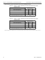
Table 2-6 Inter-Pathologist Image Analysis.
Confusion Matrix
Inter-Pathologist Image Analysis
Site 2 Site 3 Site 3
Neg Pos Neg Pos Neg Pos
47 72 38 80 38 80
Site 1
Neg 16 16 0 16 0
Pos 103 31 72 22 80
Site 2
Neg 47 36 10
Pos 72 270
% Agreement 74% 81% 90%
(95% CI) (65% - 81%) (73% - 87%) (83% - 94%)

12 Part No.: 101407300 Rev. A
Chapter 2: p53 Comparison and Reproducibility Studies p53 (DO-7) Image Analysis Software User Manual
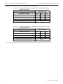
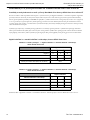
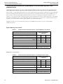
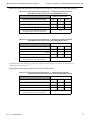
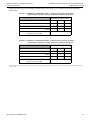
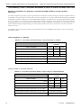
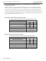
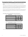
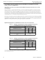
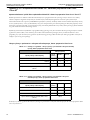
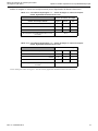
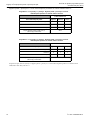
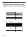
P53 Concordance with BenchMark ULTRA Instrument and iScan Coreo Slide Scanner
Study Devices and Samples for BenchMark ULTRA Instrument and iScan Coreo Slide Scanner
These p53 (DO-7) comparison studies for the Virtuoso Digital Read and Companion Algorithm Image Analysis software consisted
of 120 de-identified archived breast carcinoma sections immunohistochemically stained with CONFIRM anti-p53 (DO-7) Primary
Antibody on the BenchMark ULTRA instrument. Study test samples covered the ranges of 0-10% (negative), >10% (positive), and
were interpreted at by one pathologist All test slides were scanned at 20X magnification and all images were output in bif file format.
For the image analysis evaluation, a minimum of three fields of view were selected by each pathologist independently for each
image.120 of 120 cases were able to be evaluated and were included in the analyses below.
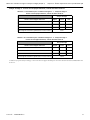
The tables below shows the concordance results for CONFIRM anti-p53 (DO-7) assay stained on the BenchMark ULTRA
instrument.
Digital Read vs. Manual
Image Analysis vs. Manual
Table 2-7 Agreement (ULTRA Instrument) - Digital Read vs. Manual
Manual Microscope Read
Digital Read
Positive Negative Total
Positive 34 14 48
Negative 0 72 72
Total 34 86 120
Positive Percent Agreement (PPA) n/N (%) (95% CI) 34/34 (100.0%) (89.8-100.0%)
Negative Percent Agreement (NPA) n/N (%) (95% CI) 72/86 (83.7%) (74.5-90.0%)
Overall Percent Agreement (OPA) n/N (%) (95% CI) 106/120 (88.3%) (81.4-92.9%)
Table 2-8 Agreement (ULTRA Instrument) - Image Analysis vs. Manual
Manual Microscope Read
Image Analysis Read
Positive Negative Total
Positive 32 4 36
Negative 2 82 84
Total 34 86 120
Positive Percent Agreement (PPA) n/N (%) (95% CI) 32/34 (94.1%) (80.9-98.4%)
Negative Percent Agreement (NPA) n/N (%) (95% CI) 82/86 (95.3%) (88.6-98.2%)
Overall Percent Agreement (OPA) n/N (%) (95% CI) 114/120 (95.0%) (89.5-97.7%)

p53 (DO-7) Image Analysis Software User Manual Chapter 2: p53 Comparison and Reproducibility Studies
Part No.: 101407300 Rev. A 13
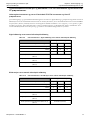
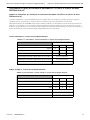
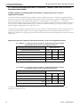
p53 Concordance with BenchMark XT/ULTRA Instruments and VENTANA iScan HT Slide
Scanner
Study Devices and Samples for BenchMark XT/ULTRA Instruments and iScan HT Slide Scanner
The p53 comparison and reproducibility studies for the Virtuoso Digital Read and Companion Algorithm image analysis software
consisted of 30 de-identified archived breast carcinoma sections immunohistochemically stained with CONFIRM anti-p53 (DO-7)
Primary Antibody. Study test samples covered the ranges of 0-10% (negative), >10% (positive), and were interpreted at by one
pathologist. All test slides were scanned at 20X magnification and all images were output in bif file format. For the image analysis
evaluation, a minimum of three fields of view were selected by each pathologist independently for each image.
Digital Read vs. Manual Microscopic Read
Image Analysis vs. Manual Microscopic Read
Table 2-9 Agreement - Digital Read vs. Manual Microscopic Read
Digital Read
Manual Microscope Read
Negative Positive Total
Negative 16 1 17
Positive 3 10 13
Total 19 11 30
Positive Percent Agreement (PPA) n/N (%) (95% CI) 10/13 (77%) (50-92%)
Negative Percent Agreement (NPA) n/N (%) (95% CI) 16/17 (94%) (73-99%)
Overall Percent Agreement (OPA) n/N (%) (95% CI) 26/30 (87%) (70-95%)
Table 2-10 Agreement - Image Analysis vs. Manual Microscopic Read
Image Analysis Read
Manual Microscope Read
Negative Positive Total
Negative 15 2 17
Positive 2 11 13
Total 17 13 30
Positive Percent Agreement (PPA) n/N (%) (95% CI) 11/13 (85%) (58-96%)
Negative Percent Agreement (NPA) n/N (%) (95% CI) 15/17 (88%) (66-97%)
Overall Percent Agreement (OPA) n/N (%) (95% CI) 26/30 (87%) (70-95%)

14 Part No.: 101407300 Rev. A
Chapter 2: p53 Comparison and Reproducibility Studies p53 (DO-7) Image Analysis Software User Manual
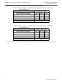
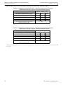
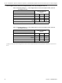
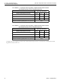
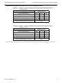
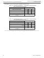
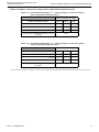
p53 Concordance with BenchMark GX Instrument and iScan Coreo and iScan HT Slide
Scanners
Study Devices and Samples for BenchMark GX Instrument and iScan Coreo and iScan HT Slide Scanners
The p53 comparison and reproducibility studies for the Virtuoso Digital Read and Companion Algorithm image analysis software
consisted of 30 de-identified archived breast carcinoma sections immunohistochemically stained with CONFIRM anti-p53 (DO-7)
Primary Antibody. Study test samples covered the ranges of 0-10% (negative), >10% (positive), and were interpreted by two
pathologists. All test slides were scanned at 20X magnification and all images were output in bif file format. For the image analysis
evaluation, a minimum of three fields of view were selected by each pathologist independently for each image.
The results were analyzed for each pathologist individually and also averaged across the two pathologists. The average overall percent
agreement (OPA) was determined as the weighted average for the two pathologists who independently scored the same cases, where
the weight of a pathologist's individual OPA rate was determined by the number of cases considered evaluable by that pathologist.
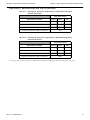
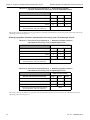
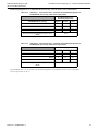
Digital Read vs. Manual Microscopic Read, iScan Coreo Slide Scanner
The average OPA for Digital Read vs. Manual Microscopic Read for iScan Coreo slide scanner was 83.3%.
Table 2-11 Pathologist #1 Agreement - Digital Read vs. Manual, iScan Coreo Slide Scanner
Digital Read
Manual Microscope Read
Negative Positive Total
Negative 17 6 23
Positive 0 7 7
Total 17 13 30
Overall Percent Agreement (OPA) n/N (%) (95% CI) 80.0%
Table 2-12 Pathologist #2 Agreement - Digital Read vs. Manual Microscopic Read,
iScan Coreo Slide Scanner
Digital Read
Manual Microscope Read
Negative Positive Total
Negative 19 2 21
Positive 2 7 9
Total 21 9 30
Overall Percent Agreement (OPA) n/N (%) (95% CI) 86.7%
A página está carregando...
A página está carregando...
A página está carregando...
A página está carregando...
A página está carregando...
A página está carregando...
A página está carregando...
A página está carregando...
A página está carregando...
A página está carregando...
A página está carregando...
A página está carregando...
A página está carregando...
A página está carregando...
A página está carregando...
A página está carregando...
A página está carregando...
A página está carregando...
A página está carregando...
A página está carregando...
A página está carregando...
A página está carregando...
A página está carregando...
A página está carregando...
A página está carregando...
A página está carregando...
A página está carregando...
A página está carregando...
A página está carregando...
A página está carregando...
A página está carregando...
A página está carregando...
A página está carregando...
A página está carregando...
A página está carregando...
A página está carregando...
A página está carregando...
A página está carregando...
A página está carregando...
A página está carregando...
A página está carregando...
A página está carregando...
A página está carregando...
A página está carregando...
A página está carregando...
A página está carregando...
A página está carregando...
A página está carregando...
A página está carregando...
A página está carregando...
A página está carregando...
A página está carregando...
A página está carregando...
A página está carregando...
A página está carregando...
A página está carregando...
A página está carregando...
A página está carregando...
A página está carregando...
A página está carregando...
A página está carregando...
A página está carregando...
A página está carregando...
A página está carregando...
A página está carregando...
A página está carregando...
A página está carregando...
A página está carregando...
A página está carregando...
A página está carregando...
A página está carregando...
A página está carregando...
A página está carregando...
A página está carregando...
A página está carregando...
A página está carregando...
A página está carregando...
A página está carregando...
A página está carregando...
A página está carregando...
A página está carregando...
A página está carregando...
A página está carregando...
A página está carregando...
A página está carregando...
A página está carregando...
A página está carregando...
A página está carregando...
A página está carregando...
A página está carregando...
A página está carregando...
A página está carregando...
A página está carregando...
A página está carregando...
A página está carregando...
A página está carregando...
A página está carregando...
A página está carregando...
A página está carregando...
A página está carregando...
A página está carregando...
A página está carregando...
A página está carregando...
A página está carregando...
A página está carregando...
A página está carregando...
A página está carregando...
A página está carregando...
A página está carregando...
A página está carregando...
A página está carregando...
A página está carregando...
A página está carregando...
A página está carregando...
A página está carregando...
A página está carregando...
A página está carregando...
A página está carregando...
A página está carregando...
A página está carregando...
A página está carregando...
A página está carregando...
A página está carregando...
A página está carregando...
A página está carregando...
A página está carregando...
A página está carregando...
A página está carregando...
A página está carregando...
A página está carregando...
A página está carregando...
A página está carregando...
A página está carregando...
A página está carregando...
A página está carregando...
A página está carregando...
A página está carregando...
A página está carregando...
A página está carregando...
A página está carregando...
A página está carregando...
A página está carregando...
A página está carregando...
A página está carregando...
A página está carregando...
A página está carregando...
A página está carregando...
A página está carregando...
A página está carregando...
A página está carregando...
A página está carregando...
A página está carregando...
A página está carregando...
A página está carregando...
A página está carregando...
A página está carregando...
A página está carregando...
A página está carregando...
A página está carregando...
A página está carregando...
A página está carregando...
A página está carregando...
A página está carregando...
A página está carregando...
A página está carregando...
A página está carregando...
A página está carregando...
A página está carregando...
A página está carregando...
A página está carregando...
A página está carregando...
A página está carregando...
A página está carregando...
A página está carregando...
A página está carregando...
A página está carregando...
A página está carregando...
A página está carregando...
A página está carregando...
A página está carregando...
A página está carregando...
A página está carregando...
A página está carregando...
A página está carregando...
A página está carregando...
A página está carregando...
A página está carregando...
A página está carregando...
A página está carregando...
A página está carregando...
A página está carregando...
A página está carregando...
A página está carregando...
A página está carregando...
A página está carregando...
A página está carregando...
A página está carregando...
A página está carregando...
A página está carregando...
A página está carregando...
A página está carregando...
A página está carregando...
A página está carregando...
A página está carregando...
A página está carregando...
A página está carregando...
A página está carregando...
A página está carregando...
A página está carregando...
A página está carregando...
A página está carregando...
A página está carregando...
A página está carregando...
A página está carregando...
A página está carregando...
A página está carregando...
A página está carregando...
A página está carregando...
A página está carregando...
A página está carregando...
A página está carregando...
A página está carregando...
A página está carregando...
A página está carregando...
A página está carregando...
A página está carregando...
A página está carregando...
A página está carregando...
A página está carregando...
A página está carregando...
A página está carregando...
A página está carregando...
A página está carregando...
A página está carregando...
A página está carregando...
A página está carregando...
A página está carregando...
A página está carregando...
A página está carregando...
A página está carregando...
A página está carregando...
A página está carregando...
A página está carregando...
A página está carregando...
A página está carregando...
A página está carregando...
A página está carregando...
A página está carregando...
A página está carregando...
A página está carregando...
A página está carregando...
A página está carregando...
A página está carregando...
A página está carregando...
A página está carregando...
A página está carregando...
A página está carregando...
A página está carregando...
A página está carregando...
A página está carregando...
A página está carregando...
A página está carregando...
A página está carregando...
A página está carregando...
A página está carregando...
A página está carregando...
A página está carregando...
A página está carregando...
A página está carregando...
A página está carregando...
A página está carregando...
A página está carregando...
A página está carregando...
A página está carregando...
A página está carregando...
A página está carregando...
A página está carregando...
A página está carregando...
A página está carregando...
A página está carregando...
A página está carregando...
A página está carregando...
A página está carregando...
A página está carregando...
A página está carregando...
A página está carregando...
A página está carregando...
A página está carregando...
A página está carregando...
A página está carregando...
A página está carregando...
A página está carregando...
A página está carregando...
A página está carregando...
A página está carregando...
A página está carregando...
A página está carregando...
A página está carregando...
A página está carregando...
A página está carregando...
A página está carregando...
A página está carregando...
A página está carregando...
A página está carregando...
A página está carregando...
A página está carregando...
A página está carregando...
A página está carregando...
A página está carregando...
A página está carregando...
A página está carregando...
A página está carregando...
A página está carregando...
A página está carregando...
A página está carregando...
A página está carregando...
A página está carregando...
A página está carregando...
A página está carregando...
A página está carregando...
A página está carregando...
A página está carregando...
A página está carregando...
A página está carregando...
A página está carregando...
A página está carregando...
A página está carregando...
A página está carregando...
A página está carregando...
A página está carregando...
A página está carregando...
A página está carregando...
A página está carregando...
A página está carregando...
A página está carregando...
A página está carregando...
A página está carregando...
A página está carregando...
A página está carregando...
A página está carregando...
A página está carregando...
A página está carregando...
A página está carregando...
A página está carregando...
A página está carregando...
A página está carregando...
A página está carregando...
A página está carregando...
A página está carregando...
A página está carregando...
A página está carregando...
A página está carregando...
A página está carregando...
A página está carregando...
A página está carregando...
A página está carregando...
A página está carregando...
A página está carregando...
A página está carregando...
A página está carregando...
A página está carregando...
A página está carregando...
A página está carregando...
A página está carregando...
A página está carregando...
A página está carregando...
A página está carregando...
A página está carregando...
A página está carregando...
A página está carregando...
-
 1
1
-
 2
2
-
 3
3
-
 4
4
-
 5
5
-
 6
6
-
 7
7
-
 8
8
-
 9
9
-
 10
10
-
 11
11
-
 12
12
-
 13
13
-
 14
14
-
 15
15
-
 16
16
-
 17
17
-
 18
18
-
 19
19
-
 20
20
-
 21
21
-
 22
22
-
 23
23
-
 24
24
-
 25
25
-
 26
26
-
 27
27
-
 28
28
-
 29
29
-
 30
30
-
 31
31
-
 32
32
-
 33
33
-
 34
34
-
 35
35
-
 36
36
-
 37
37
-
 38
38
-
 39
39
-
 40
40
-
 41
41
-
 42
42
-
 43
43
-
 44
44
-
 45
45
-
 46
46
-
 47
47
-
 48
48
-
 49
49
-
 50
50
-
 51
51
-
 52
52
-
 53
53
-
 54
54
-
 55
55
-
 56
56
-
 57
57
-
 58
58
-
 59
59
-
 60
60
-
 61
61
-
 62
62
-
 63
63
-
 64
64
-
 65
65
-
 66
66
-
 67
67
-
 68
68
-
 69
69
-
 70
70
-
 71
71
-
 72
72
-
 73
73
-
 74
74
-
 75
75
-
 76
76
-
 77
77
-
 78
78
-
 79
79
-
 80
80
-
 81
81
-
 82
82
-
 83
83
-
 84
84
-
 85
85
-
 86
86
-
 87
87
-
 88
88
-
 89
89
-
 90
90
-
 91
91
-
 92
92
-
 93
93
-
 94
94
-
 95
95
-
 96
96
-
 97
97
-
 98
98
-
 99
99
-
 100
100
-
 101
101
-
 102
102
-
 103
103
-
 104
104
-
 105
105
-
 106
106
-
 107
107
-
 108
108
-
 109
109
-
 110
110
-
 111
111
-
 112
112
-
 113
113
-
 114
114
-
 115
115
-
 116
116
-
 117
117
-
 118
118
-
 119
119
-
 120
120
-
 121
121
-
 122
122
-
 123
123
-
 124
124
-
 125
125
-
 126
126
-
 127
127
-
 128
128
-
 129
129
-
 130
130
-
 131
131
-
 132
132
-
 133
133
-
 134
134
-
 135
135
-
 136
136
-
 137
137
-
 138
138
-
 139
139
-
 140
140
-
 141
141
-
 142
142
-
 143
143
-
 144
144
-
 145
145
-
 146
146
-
 147
147
-
 148
148
-
 149
149
-
 150
150
-
 151
151
-
 152
152
-
 153
153
-
 154
154
-
 155
155
-
 156
156
-
 157
157
-
 158
158
-
 159
159
-
 160
160
-
 161
161
-
 162
162
-
 163
163
-
 164
164
-
 165
165
-
 166
166
-
 167
167
-
 168
168
-
 169
169
-
 170
170
-
 171
171
-
 172
172
-
 173
173
-
 174
174
-
 175
175
-
 176
176
-
 177
177
-
 178
178
-
 179
179
-
 180
180
-
 181
181
-
 182
182
-
 183
183
-
 184
184
-
 185
185
-
 186
186
-
 187
187
-
 188
188
-
 189
189
-
 190
190
-
 191
191
-
 192
192
-
 193
193
-
 194
194
-
 195
195
-
 196
196
-
 197
197
-
 198
198
-
 199
199
-
 200
200
-
 201
201
-
 202
202
-
 203
203
-
 204
204
-
 205
205
-
 206
206
-
 207
207
-
 208
208
-
 209
209
-
 210
210
-
 211
211
-
 212
212
-
 213
213
-
 214
214
-
 215
215
-
 216
216
-
 217
217
-
 218
218
-
 219
219
-
 220
220
-
 221
221
-
 222
222
-
 223
223
-
 224
224
-
 225
225
-
 226
226
-
 227
227
-
 228
228
-
 229
229
-
 230
230
-
 231
231
-
 232
232
-
 233
233
-
 234
234
-
 235
235
-
 236
236
-
 237
237
-
 238
238
-
 239
239
-
 240
240
-
 241
241
-
 242
242
-
 243
243
-
 244
244
-
 245
245
-
 246
246
-
 247
247
-
 248
248
-
 249
249
-
 250
250
-
 251
251
-
 252
252
-
 253
253
-
 254
254
-
 255
255
-
 256
256
-
 257
257
-
 258
258
-
 259
259
-
 260
260
-
 261
261
-
 262
262
-
 263
263
-
 264
264
-
 265
265
-
 266
266
-
 267
267
-
 268
268
-
 269
269
-
 270
270
-
 271
271
-
 272
272
-
 273
273
-
 274
274
-
 275
275
-
 276
276
-
 277
277
-
 278
278
-
 279
279
-
 280
280
-
 281
281
-
 282
282
-
 283
283
-
 284
284
-
 285
285
-
 286
286
-
 287
287
-
 288
288
-
 289
289
-
 290
290
-
 291
291
-
 292
292
-
 293
293
-
 294
294
-
 295
295
-
 296
296
-
 297
297
-
 298
298
-
 299
299
-
 300
300
-
 301
301
-
 302
302
-
 303
303
-
 304
304
-
 305
305
-
 306
306
-
 307
307
-
 308
308
-
 309
309
-
 310
310
-
 311
311
-
 312
312
-
 313
313
-
 314
314
-
 315
315
-
 316
316
-
 317
317
-
 318
318
-
 319
319
-
 320
320
-
 321
321
-
 322
322
-
 323
323
-
 324
324
-
 325
325
-
 326
326
-
 327
327
-
 328
328
-
 329
329
-
 330
330
-
 331
331
-
 332
332
-
 333
333
-
 334
334
-
 335
335
-
 336
336
-
 337
337
-
 338
338
-
 339
339
-
 340
340
-
 341
341
-
 342
342
-
 343
343
-
 344
344
-
 345
345
-
 346
346
-
 347
347
-
 348
348
-
 349
349
-
 350
350
-
 351
351
-
 352
352
-
 353
353
-
 354
354
-
 355
355
-
 356
356
-
 357
357
-
 358
358
-
 359
359
-
 360
360
-
 361
361
-
 362
362
-
 363
363
-
 364
364
-
 365
365
-
 366
366
-
 367
367
-
 368
368
-
 369
369
-
 370
370
-
 371
371
-
 372
372
-
 373
373
-
 374
374
-
 375
375
-
 376
376
-
 377
377
-
 378
378
-
 379
379
-
 380
380
-
 381
381
-
 382
382
-
 383
383
-
 384
384
-
 385
385
-
 386
386
-
 387
387
-
 388
388
-
 389
389
-
 390
390


em outras línguas
- italiano: Roche Virtuoso Manuale utente
- slovenčina: Roche Virtuoso Používateľská príručka
- dansk: Roche Virtuoso Brugermanual